Retinal Artery Occlusions
What are retinal vein occlusions?
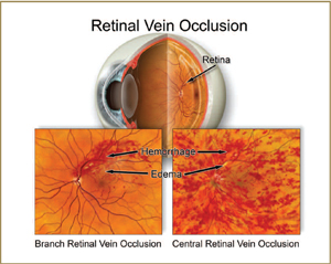
A retinal vein occlusion occurs when one of the veins of the retina becomes blocked and cannot drain blood from the retina normally. This blockage leads to a backup of blood and fluid into the retina. This back up of blood can be seen in the retina as hemorrhages. The amount of swelling in the retina varies and depends on the degree of blockage of the retinal vein.
Two types of vein occlusions
Central Retinal Vein Occlusion (CRVO)
If the blockage involves the central vein to the retina, located at the optic nerve, it is called a central retinal vein occlusion. In these cases, hemorrhages can be scattered throughout the retina, and often the central portion of the retina, the macula, can become swollen.
Branch Retinal Vein Occlusion (BRVO)
If the blockage involves only one of the smaller retinal veins, it is called a branch retinal vein occlusion. In these cases, hemorrhages and swelling are limited to the area of the retina that is drained by that particular vein.
Risk factors for vein occlusions
- High blood pressure
- Diabetes
- Age
- Blood clotting disorders
Symptoms
Symptoms of a retinal vein occlusion can vary based on the severity of the blockage in the vein and amount of swelling in the retina. Common symptoms include:
- Painless loss of vision
- Blurry vision
- In severe cases, pain from increased eye pressure
Diagnosis
Your doctor can diagnose a CRVO or BRVO when examining your eyes based on the appearance of hemorrhages and swelling in the retina. Other diagnostic tests such as fluorescein angiogram and optical coherence tomography (OCT) can be performed to examine the degree of blockage of the vein and the amount of swelling present in the retina.
Treatment
There is no cure for a vein occlusion. However, the main reason for decreased vision from a retinal vein occlusion is swelling in the central part of the retina called macular edema. Treatments for swelling include:
- Steroid injection
- Injection with medicines that decrease blood vessel leakage (Avastin, Lucentis, Macugen)
- Laser treatment
In mild vein occlusions, often there is no significant swelling, and no treatment is indicated. In severe cases of CRVO, abnormal blood vessels can develop that can lead to glaucoma. These cases are often treated with laser and/or injection with a medication to decrease blood vessel leakage. Some patients do require surgery to control eye pressure even after laser or injections.
What are retinal artery occlusions?
Retinal artery occlusions occur when a plaque or clot becomes lodged in a retinal artery and prevents blood from flowing past it. These blockages cause damage to the retina due to lack of blood flow. In the short term, this loss of blood flow can cause swelling of the retina. If the blood flow remains poor, it can lead to death of the retina cells and subsequent loss of vision.
Types of Retinal Artery Occlusion
- Central Retinal Artery Occlusion (CRAO) – A central retinal artery occlusion occurs from a blockage of the main retinal artery as it enters the eye at the optic nerve.
- Branch Retinal Artery Occlusion (BRAO) – A branch retinal artery occlusion involves blockage of one or more of the smaller retinal arteries.
Risk factors
- High blood pressure
- Cardiovascular disease
- High cholesterol
- Carotid artery disease
Symptoms
Symptoms of a retinal artery occlusion vary based on the degree of impairment of blood flow to the eye. Sudden, painless loss of vision, either partial or complete, is the most common symptom. Loss of visual field, or a blurry or gray spot in the vision can also occur.
Diagnosis
Your doctor can diagnose a retinal artery occlusion based on examination of your eyes. Often a clot or plaque can be seen inside the blocked artery. Other diagnostic tests such as a fluorescein angiogram can be helpful in determining the degree of loss of blood flow.
Often your doctor will recommend that you see your regular physician for a complete cardiovascular work-up to evaluate the risk factors listed above.
Treatment
Unfortunately, there are no good treatments for a retinal artery occlusion. In very early cases, attempts can be made to lower the eye pressure with eye drops or a procedure called anterior chamber paracentesis. However, even with eye pressure lowering treatments, there is often little change in the blood flow status of the eye. In severe cases, abnormal blood vessels can grow that cause eye pain and glaucoma to develop. In these cases, laser treatment can be used to try to help control the eye pressure and relieve pain.